Introduction
Department of Anatomy
- The Department of Anatomy aspires to become the best at teaching the structure of the human body in a clinically relevant context. The anatomical sciences are essential disciplines for understanding medicine and form the foundation for medical studies. We strive to develop and promote medical knowledge by encompassing education, instruction, research and attitudes. Our mission is to provide a strong foundation for new beginners by teaching basic science of medicine and surgery
- Anatomy department is located on the ground floor of academic block of Army college of Medical Sciences, Delhi Cantt with a team of nine faculty members. The infrastructure of Anatomy department consists of Histology Practical Lab with slide preparation room, Dissection Hall with attached bone room, Embalming room and tank storage rooms, Library cum seminar room, Lecture Hall, Demonstration rooms, Research Laboratory, Museum, Artist cum modeller room, Cold storage and Bone Saw room, and Burial ground.
- A great deal of dedication and effort has been put in at the department since its inception. Teaching of anatomy is accomplished by the way of small groups teaching and learning ,lectures and demonstrations by using audio-visual aids and student seminars .Practical training is imparted by dissection and demonstration on cadavers and prosected specimens, histology laboratory practicals and DOAP (Demonstrate, observe ,assist and perform ) sessions. Frequent assessments are conducted in the form of spotting exams, OSPE and Viva voce. Our faculty are well versed with CBME competencies and their knowledge is frequently updated by CME cum hands on workshops on medical education and guest lectures.
- Cadaveric dissection is the integral part of the undergraduate teaching of all medical students to update their dissecting skills. It helps the student to learn topographic localization of organs in the body. Our department encourages and promotes voluntary body donation programme and also provides embalming facility with well trained staff. We aspire to impart teaching and learning and produce excellence in the field of histology, osteology, gross anatomy, neuroanatomy, developmental anatomy, clinical and functional anatomy, biological anthropology and medical education. The department has adequately equipped research laboratories to conduct both applied and basic research.
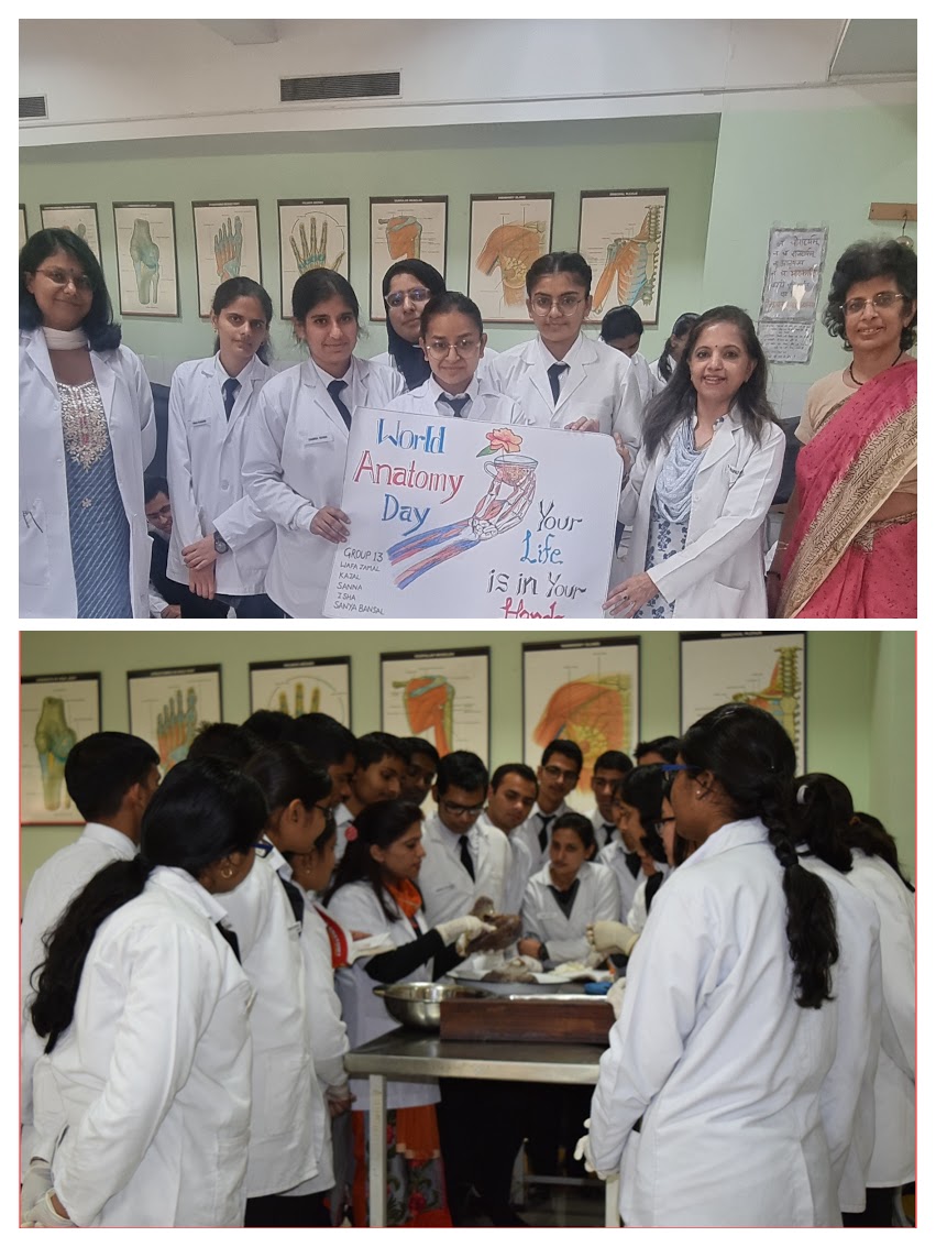
- The Anatomy department organizes a drawing competition each year on 15th October in remembrance of Andreas Vesalius, the father of anatomy in which students showcase their artistic talent by drawing and painting anatomy sketches.
- The department regularly conducts cadaveric workshops in surgical and allied disciplines to train specialist from all over India in surgical techniques. Recent workshops conducted in the department include
a. DIRECT ANTERIOR ADVANTAGE-A CME cum cadaveric workshop on Direct anterior Approach in Total Hip Replacement with R& R hospital on 22nd September 2023-12-23
b. AFCONHNS-Armed forces conference on Head & Neck cadaveric dissection with R&R Hospital & BHDC from 25th-27th May 2023

OUR DISSECTION HALL
The Dissection Hall is well-ventilated, spacious and well lit. It is fully air-conditioned with a capacity to accommodate about 150 students. It is a fully formalin free dissection hall .Our department initiated a novel method of body preservation by using a user-friendly preservative, ‘phenoxyethanol’ which is less-carcinogenic, pleasant smelling and causes less irritation, unlike formalin. In addition, separate rooms for bones, cadaver preparation, embalming, cadaver storage, cold storage, and lockers are attached to the Dissection Hall. We are fortunate to have an adequate number of donated bodies for dissection and the students are encouraged to pay respect to the ‘Cadaver- their first teacher’ and master their skill in dissection and understand the intricate human body.
On the orientation day to the anatomy department we pay homage to the cadavers along with the students and their parents by reciting a homage prayer for the departed souls who donated their bodies for medical teaching. The DH has donor boards to felicitate the noble souls who participated in the body donation program.
MUSEUM & CROSS SECTIONAL IMAGING LAB
Our Museum is located on the ground floor of the academic block. Gross anatomy section has more than 150 dissected human specimens kept in transparent containers filled with ten percent formalin. Specimen labels describe the relevant anatomy for reference. Every year we add newly dissected specimens to the collection. We have more than 180 embryology models for the students to study and correlate the fundamentals of developmental anatomy. Osteology section features disarticulated real human bones displayed region wise and articulated joints for anatomical correlation.
Cross sectional Imaging Section has view boxes in the form of tables to display the plain and contrast X-rays. Highlight of the museum are the two walls: one displays cross sectional anatomy through actual MRI images and the other displays high contrast histology images. Each image in the cross sectional anatomy has 4 parts: a diagram showing the level at which section is taken; gross anatomy cross section or longitudinal section from that level; MRI image from the same level; hand drawn diagram of the gross anatomy section with labels. This makes it easier for the learner to correlate the gross anatomy appearance with the radiological image and enhances cognition. The high contrast histology images displays well labelled 85 tissue slides for recall and revisions.
HISTOLOGY LABORATORY
Our Histology lab is equipped with more than 60 monocular & four binocular microscopes, charts, Slides and LCD Projector. We have a trinocular display microscope fitted with a camera and augmented with a visual display unit for understanding of histology slides. Our departmental library has about 300 books of all subdivisions of Anatomy with recent editions and new texts are regularly updated. The department is also having two demonstration rooms for small group teaching and demonstrations and is equipped with mobile and enclosed skeletons, computer and LCD projector. The Research Lab is equipped with anthropometric instruments, automatic tissue processor and microtome.

 Faculty Details